Rajkot
+919405000222

'inverted microscope for metallography analysis'

Items tagged with 'inverted microscope for metallography analysis'

product image
Metallurgical Microscopes

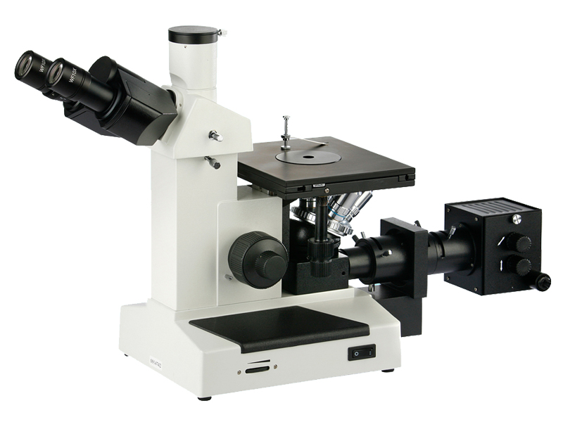
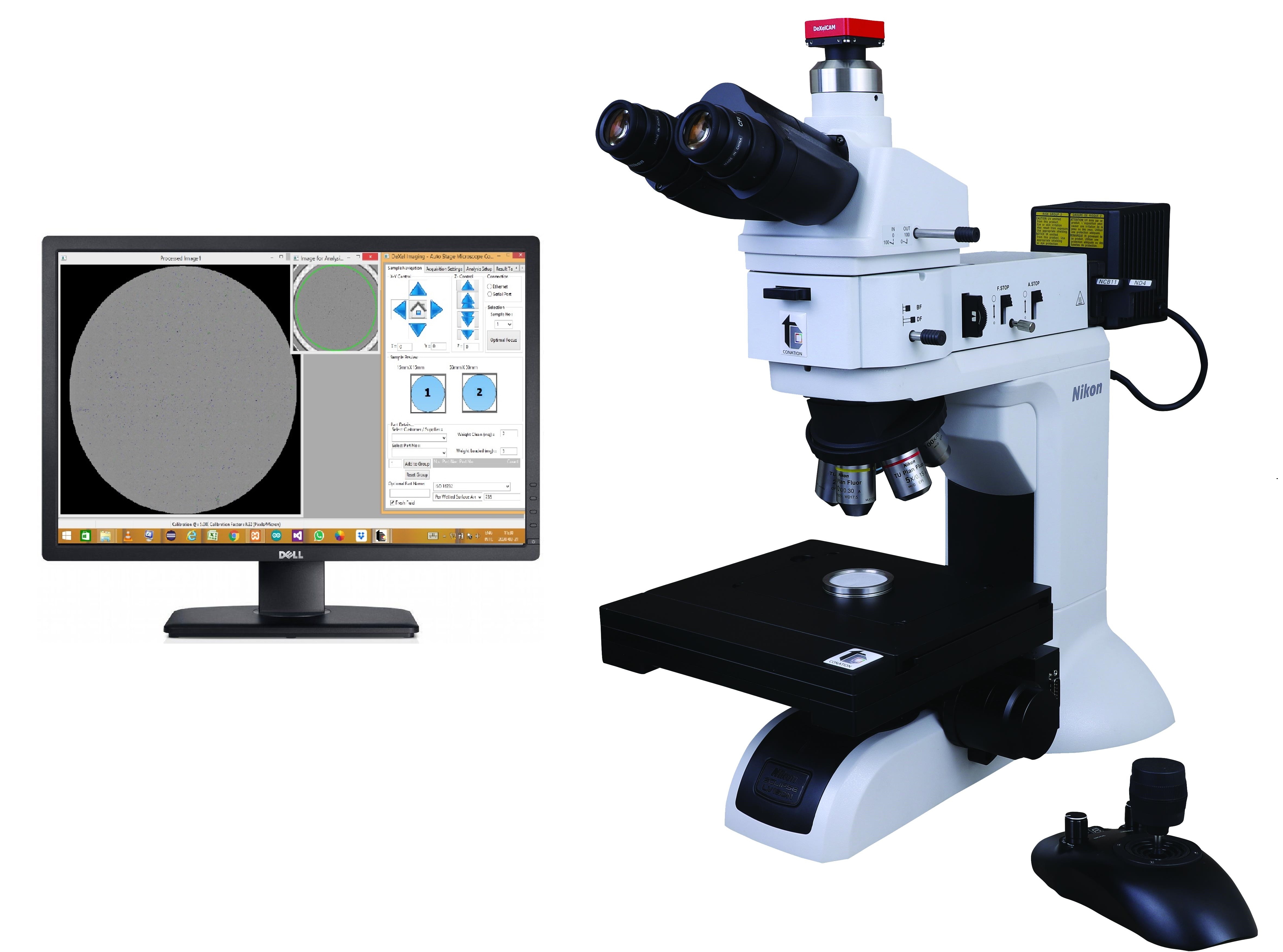
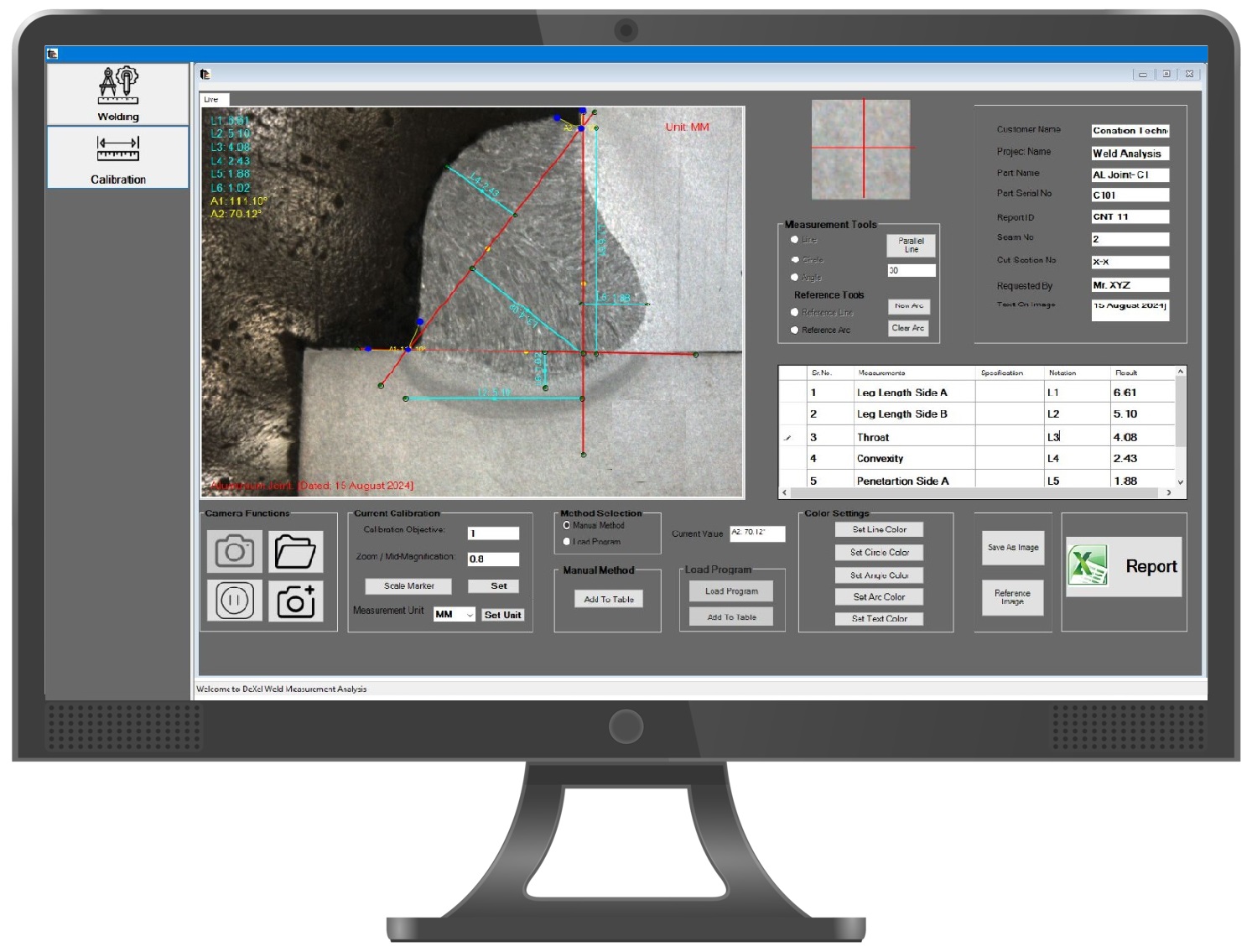
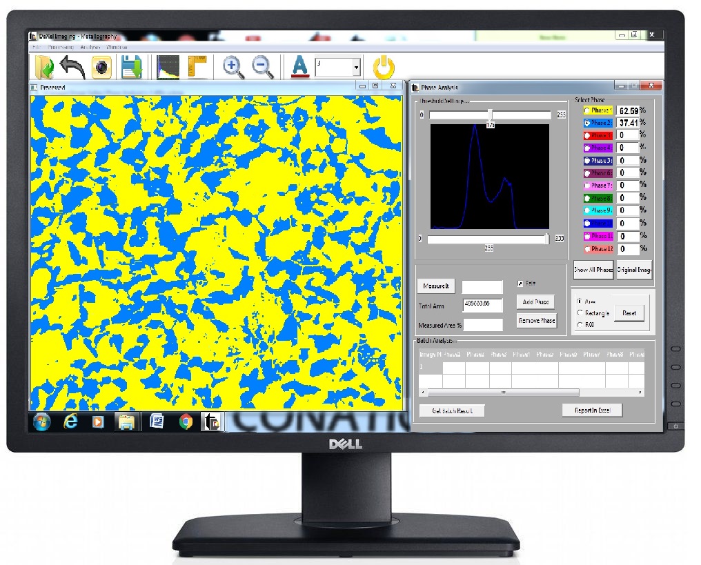
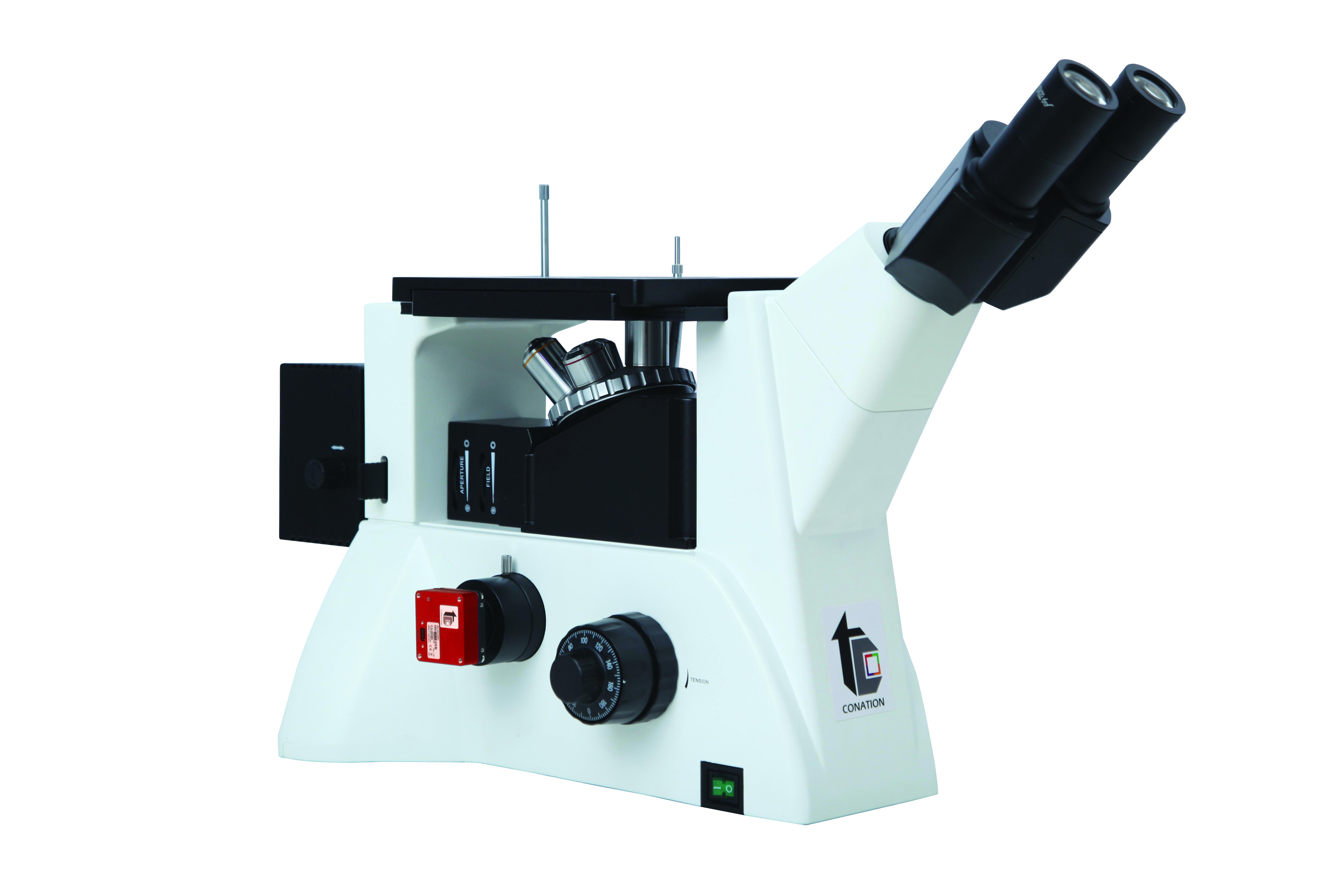
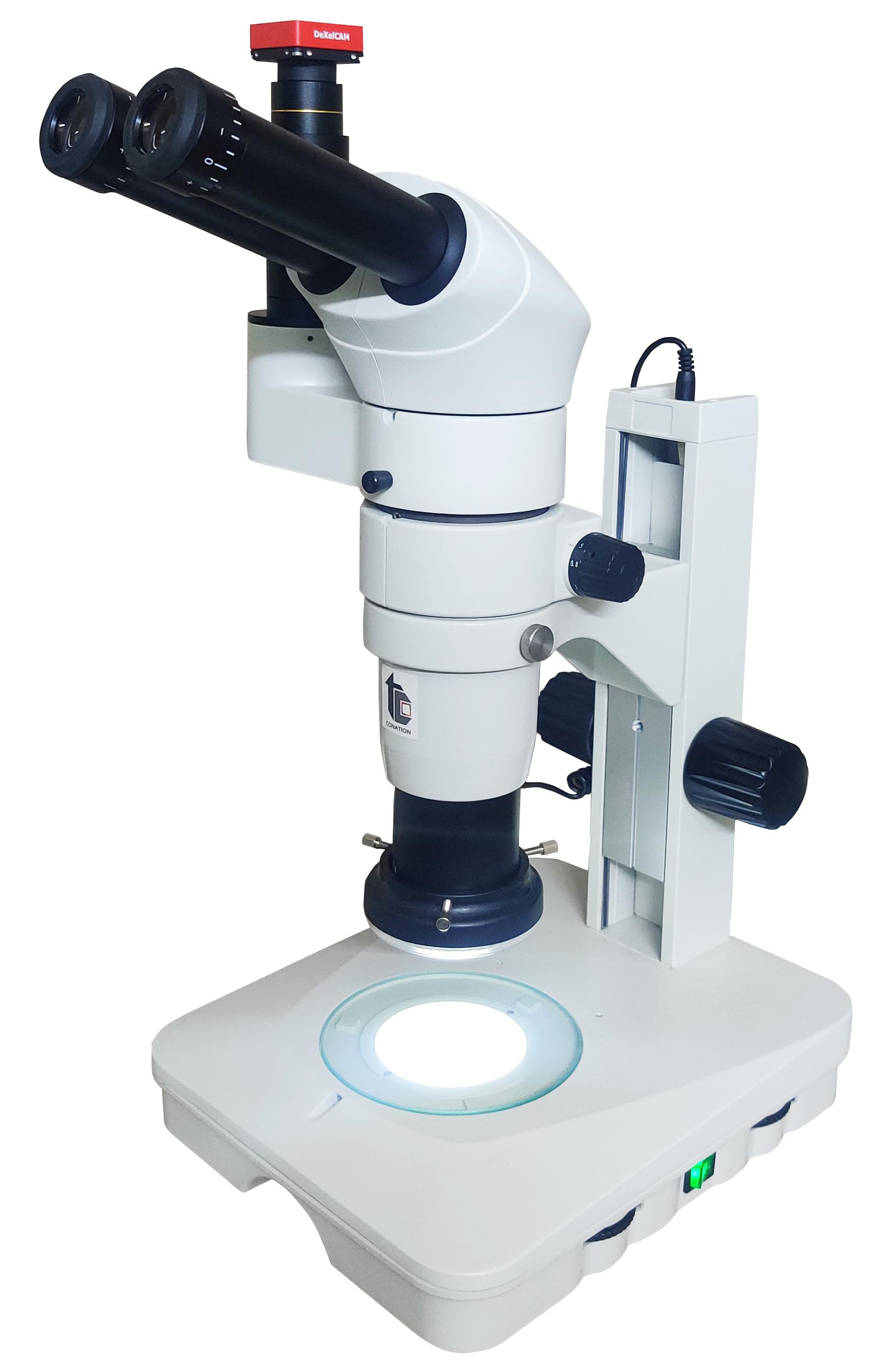
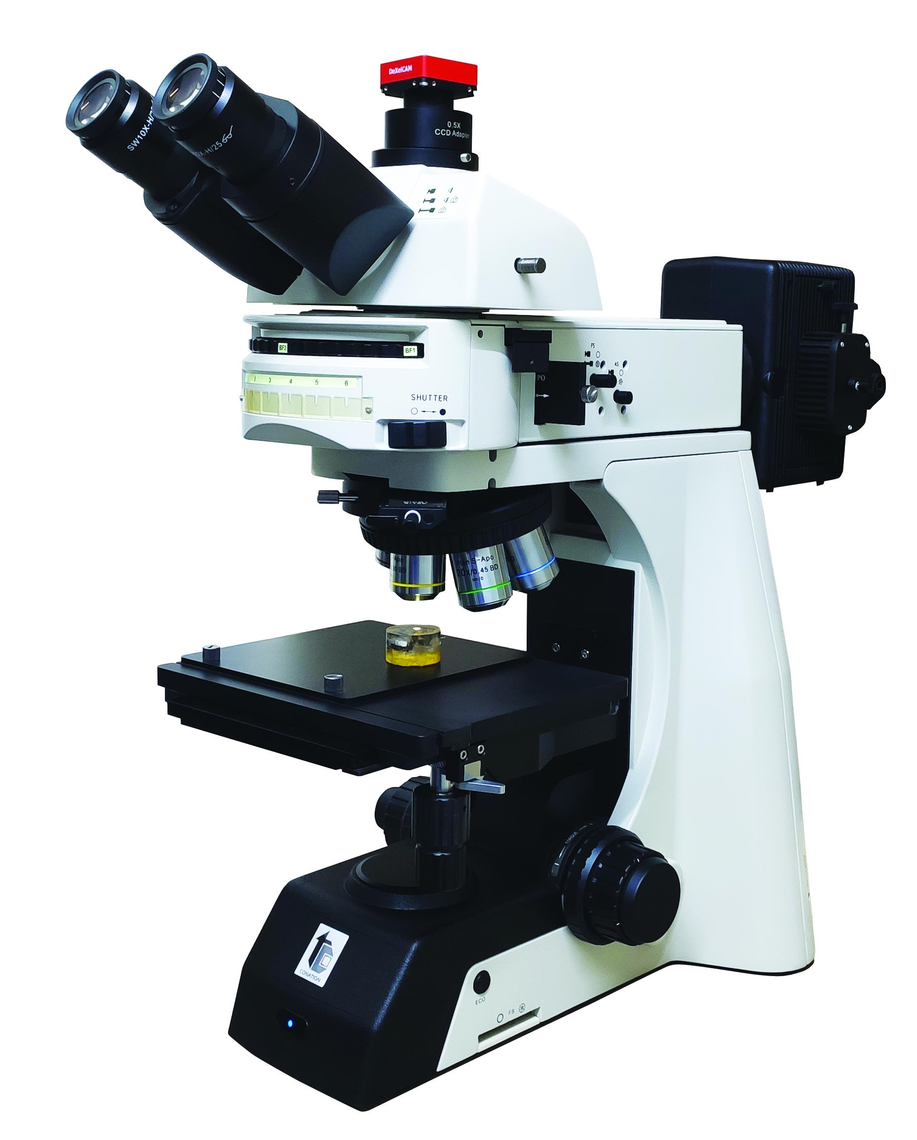
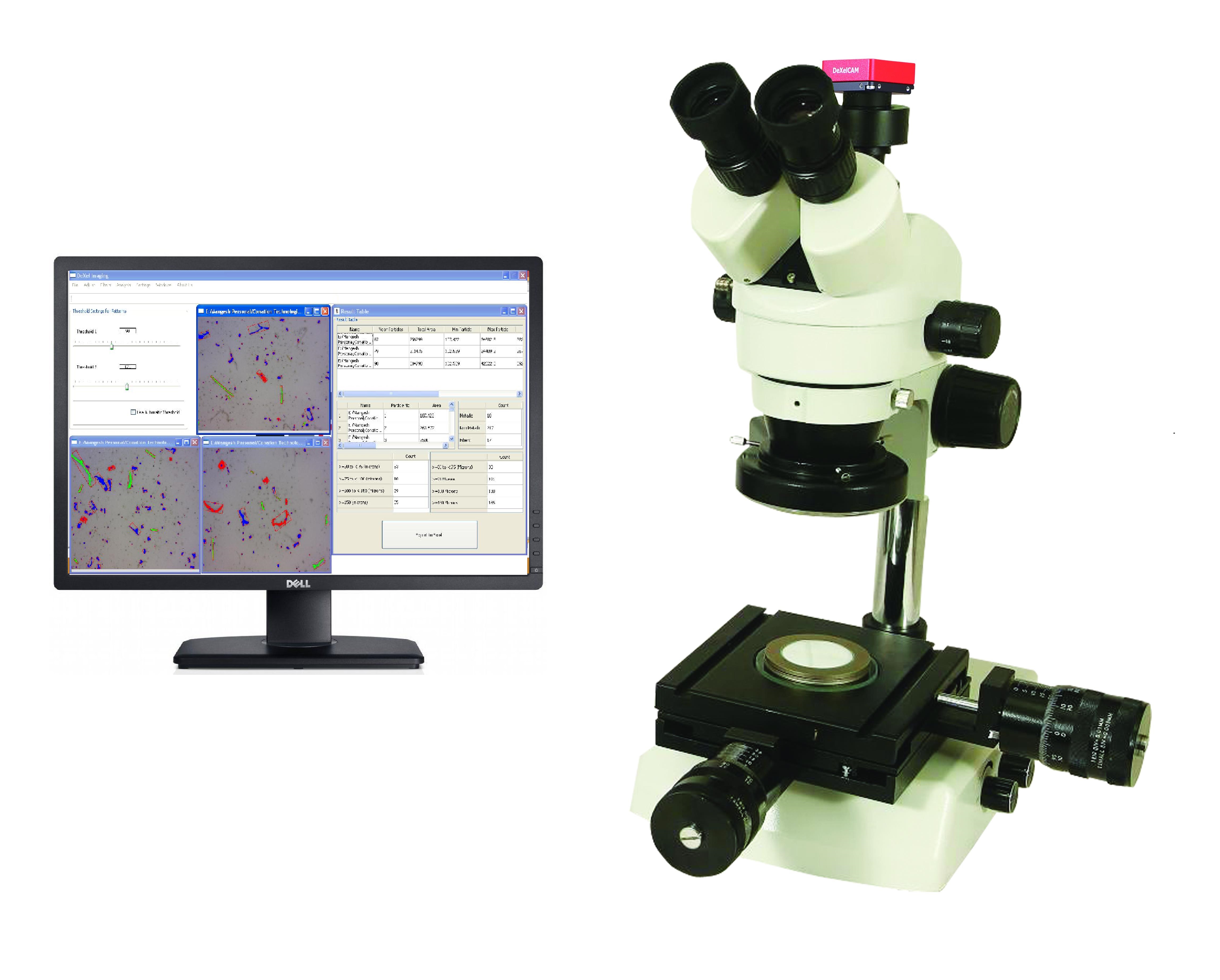
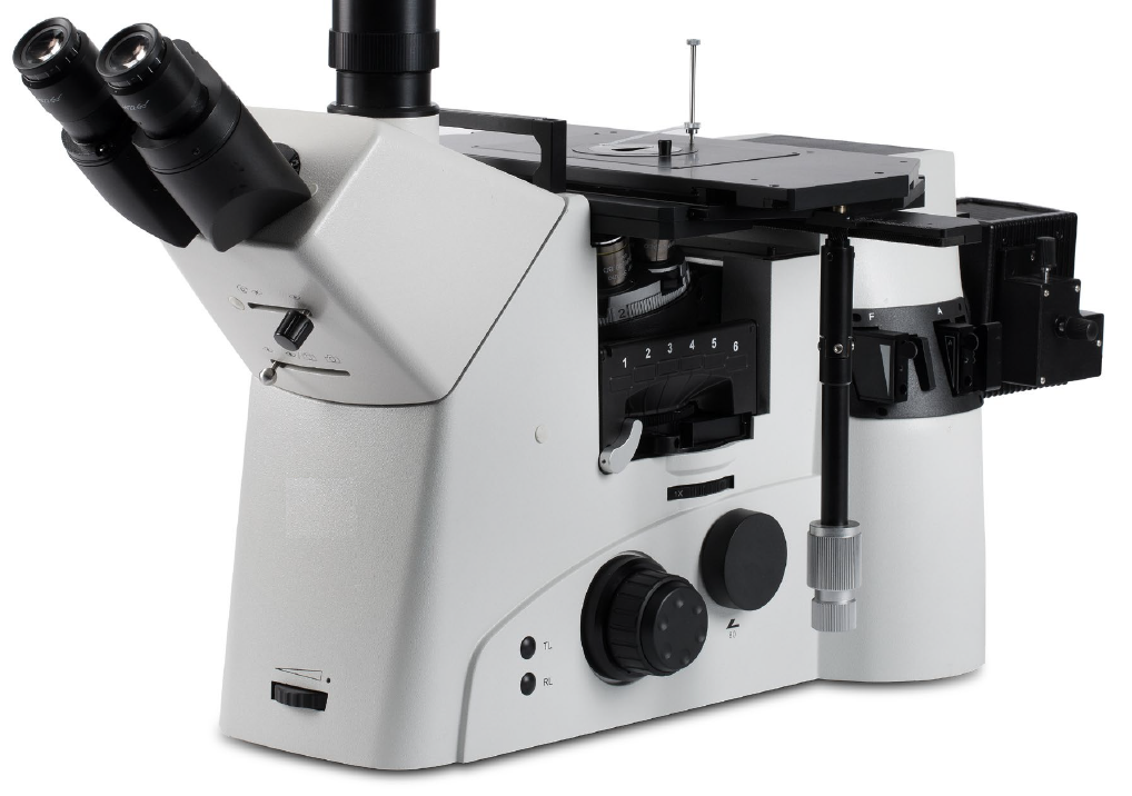
This is top of the class material microscopes that incorporated semi-apochromat and Apo-chromat objectives. These objectives have exceptionally high numerical aperture that give best possible resolution in optical microscopy along with superior clarity and sharpness. Microscopes has modular design to offer greater flexibility to adapt various observation modes and extended magnification with intermediate magnification changer. Highlights • Excellent resolution right upto 1500X with high NA objectives and intermediate magnification changer • Semi-apochromat and Apo-chromat objectives with exceptionally high numerical aperture to give best possible resolution, high contrast and sharp images • Observations modes – Brightfield, Darkfield, Polarizer; Option for DIC • Large sample stage with larger XY moving range • Sturdy structure with optimized design for extended life span Specifications Body - Inverted type Observation modes - Brightfield, Darkfield, Polarizer Illuminations - 12V, 100W Halogen / LED Filters • Integrated field diaphragm, aperture diaphragm • ND6/ND25 filter, Green filter, Blue filter Eyepiece tube - Trinocular, inclined 45˚; diopter adjustment (±5) with an interpupillary distance adjustment range of 55 to 75mm Eyepieces - 10X wide field, FOV Φ 22 mm Nosepiece - Sextuple nosepiece (DIC slot) Objectives - Infinity plan achromatic objectives • 5X BD Semi-Apo; 10X BD Semi-Apo; 20X BD Semi-Apo: 50X BD APO; 100X BD APO Polarizer Analyzer and polarizer Focusing Mechanism - Coaxial coarse and fine focusing controls, Adjustable tension control; Fine division 1 µm; Moving range 9mm, up 7mm and down 2mm Stage - Size 340 mm x 230 mm; Travel area 130 mm x 85 mm Optional Features - DIC and fluorescent Attachment; Ergo tilting trinocular head; LED illumination; Stage plates Attachments for Inverted Microscopes • Digital Color Cameras – Ranging from 1.3 to 20 MP resolution; CCD/CMOS; USB 2.0/USB 3.0/Ethernet connectivity • Metallography Image Analyzer - Comprehensive software for all metallography applications

Send Message
product image
Metallurgical Microscopes

Microscope with capability of 1000X magnification with plan achromatic lenses This is basic Inverted Microscope specially designed for routine applications. Being very cost effective yet with high resolution, they are ideally suitable for forging and foundry applications, and educational purpose. • Observation – Brightfield; Polarizer optional • Eyepiece Tube - Trinocular (seidentopf) head, inclined at 30 deg, rotates full 360 deg., with built-in C-mount tube • Eyepiece – WF 10X/18 • Objectives - DIN plan achromat; corrected for spherical and color aberration; PL 10X, 20X, 40X, 100X. (Optional 5X, 50X, 60X, 80X) • Nosepiece - Quadruple/Quituple, ball bearing nosepiece. • Illumination – LED illumination / Khohler 6V/20W halogen illumination; full range intensity variation, filters included • Focusing - Coaxial course and fine focusing with adjustable tension control. • Stage - 150mmX185mm, travel range - 15mmX15mm, low position right hand control.

Send Message
product image
Cleanliness Particle Analyzers

Critical components going into fluid or air circuitry, engine components and some trasmission assemblies are need to free from particle contamination arising from manufacturing and assembly process. Particle contamination beyond limit will invariably affect the performance of the component during it’s life cycle. Conation Technologies, a pioneer in India for cleanliness test equipment manufacturing, offers range of Automatic Particle Analysis Systems. Systems are compliant to most of the international standards such as VDA19.1, ISO16232-2018, ISO 4406 & 4407, ASTM F312 etc Applications • Engine, Transmission, Braking components • Fuel and hydraulic line components • Electronic components • Fluids – Fuel, oils, coolants etc • Cleanliness Analysis as per VDA19.1, ISO16232-2018, ISO 4406 & 4407, ASTM F312 Features/Highlights • Particle size measurement right from 1 micron • With Nikon’s unparalleled optics, each particle is captured with high resolution and measured to the highest accuracy. • Quick scanning and faster processing to deliver the report in short time • High resolution German made camera • High precision ultra compact motorized stage with XYZ automation; programmable • State of the art, Unified and simplified user interface software; user friendly navigation • Intelligent Metallic Particle separation • Advanced Fiber Identification with Elongated Length feature • Adaptability for range of filters/membranes such as cellulose, Nylon, PTFE etc; • Options for 25 mm and 90 mm scanning also available • Specially designed Database and Reporting software Specifications Nikon Microscope; Model: LV150N • Modularized LV150 series Microscope with CFI60 Optical System • CFI TU Plan FLUOR Epi 5X, 10X and 20X with high NA objectives • Trinocular eyepiece tube ESD • CFI 10X with diopter adjustment (F.O.V. 22mm) • LED illumination with internal power source and intensity control, field diaphragm and aperture stop, can accept filters • Sextuple Nosepiece • Polarizer and Analyzer • Expandable for Darkfield and DIC observation modes  World Class Nikon optics with Superior Optical performance and great resolution to visualize and capture crisp and clear images  Best in class with Higher NA  Highly corrected for chromatic aberration Ultra compact Motorized stage with controller and Joystick • High Precision screw and stepper motor driven XY stage • Travel range - 80X60 mm • Positioning accuracy - 10 micron • Motorized Focusing (Z Drive) with adapter • Controller for motorized X, Y and Z operation • Joystick to operate motorized stage • Specially designed sample holder for 47mm dia filter paper High resolution camera, Made in Germany • USB2.0, CMOS, Color • 5 MP, High resolution • 2.2X2.2 square pixel • Rolling/Global shutter Imaging Software  DeXel-Filtrate Imaging software • Unified user interface software to harmonize the operation across all automatic models • Specially designed Image analyzer to analyze and measure all particles on filter paper as per any applicable standard such as ISO16232-18, VDA19.1, ISO4406 etc • Stitch all captured images to generate a single image of entire filter paper; • Automatically analyze this single image for determination of particulate contamination as per desired standard • Separation of particles as Metallic, Non-metallic, and fibres • Distribution all particles into user/standard defined size classes • Estimation of Filter Occupancy (Patch Saturation) • Real time Auto-Focusing • Intelligent Metallic Particle separation • Advanced Fiber Identification with Elongated Length feature  Stage Operating Software • XYZ movement control of stage from PC

Send Message
product image
Image Analyzers

Weld structure analysis is extremely important to understand the quality of the weld, which in turn helps to understand the strength, performance and durability of the welded component. Any kind of weld such as butt weld, lap weld or T-joint weld should pass through the stringent macrographic inspection to reveal the intended quality, anomalies or disparities. Similar to metallography analysis, weld joint need to be cut in cross-sectional direction, polished to the mirror finish and etched with appropriate etchant to reveal the structure of bead, surrounding area, heat affected zone, weld passes etc. This sample then is placed under stereozoom microscope and further analysis can be carried out with DeXel-Weld Image analyzer. Weld Joint programs – Software has preloaded list with measurement parameters specific to type of weld joint like butt weld, lap weld, T-weld, corner weld, edge weld etc. Extensive list of parameters – List includes all the geometric parameters of weld such as Leg lengths, throat, penetrations, root penetration, metal thickness, gap, Area and depth of Heat Affected Zone, Joining Angles etc Macro based analysis - User can also preselect the parameters from the list and create a customized list depending upon specific requirements. The best part is that the macro can be created in user-friendly excel format. Color coded Measurements - All the measurements are color coded for better and quick understanding of the report. Porosity and other defect Analysis - Geometrical parameters such as length, width, area, perimeter estimation of defects such as porosity, crack, lack of fusion etc 2D analysis of features - Object counting, distance between any two points, angle between any two edges, Area of circle rectangle, irregular shape etc. OK/NOK Report – Limit values (Acceptance specifications) can be assigned to the each parameter and derived value can be compared which immediately confirms OK or Not OK reading. Report Generation – Simplified report in predefined excel template with facility to add logo, address etc.

Send Message
product image
Image Analyzers

Qualification and quantification of microstructural features have always been crux of any metallurgical Analysis. Digitization of the microstructure with the help of image capturing hardware and storage into computer, analysis of these images with specialized image processing & analysis software modules has changed the complete scenario in material characterization. Tremendous advancements in computer technology and software developments has shifted manual interpretation methods to computerized ones in metallography, just like any other field. Image analyzers (comprehensive term which include image capturing, processing, analysis and reporting) has given great advantage to metallurgists in their everyday activities as well as in their research. Fatigue-free and User-friendly Analysis of microstructure with specialized image analyzers relieves tediousness and fatigue associated with manual measurements. Analyzing hundreds of fields manually could create tremendous fatigue and force the errors during analysis which are completely avoided in analysis through Image analyzers. Fast Just like any other computerized application, analysis of microstructure can be done extremely fast with image analyzers which also increases the productivity. Precision and Accuracy Inherent nature of software coding which is implementation of mathematical formulae renders highest possible precision and accuracy in any of the microstructure analysis. Repeatability and Reproducibility Computer being a deterministic machine which will always yield same results when same program is run on the same input. This makes Image Analyzer give full repeatability and reproducibility. Data Preservation All the analyses done and reports generated can be stored in digital format in your computer and you can retrieve the same any time in future without any much hassle. Dependability Though Image analyzer cannot replace the metallurgist, however a well trained staff can perform highly dependable analysis and produce the reliable results. Compliance to standards Mathematical and non-mathematical inputs from various ASTM, JIS, DIN and other international standards are typically translated into programs while writing the software of Image analyzer. So eventually these software modules are compliant to these standards. Cost Efficient After the initial cost of investment is done, there is no running cost for Image analyzer. This makes their use extremely cost efficient in daily use. -------------------------------------------------------------------------------------------------------------------------------------------------- Applications -Raw Material -Steel Mills -Forgings -Ferrous and Non-ferrous Castings -Heat Treatment -Weld structures -Electronic components -Petrography -Cement and Clinker -Material science Key Features Application Specific Analysis Modules – Grain size measurement; Phase analysis; inclusion analysis; nodularity, nodule count and size classification; graphite flake type, count and size classification; Porosity measurement, Banding Analysis, Decarb and coating thickness and many more… 2D analysis – Modules for measurement of count and various geometric aspects of features such as length, width, area, parameter, angle etc

Send Message
product image
Metallurgical Microscopes

These microscopes provides superior clarity and sharpness due its infinity optical system and high numercial aperture objectives. Highlights • Excellent resolution right upto 1000X with high NA objectives • High contrast, sharp images • Versatile in nature • Illumination Options – Brightfield, Darkfield, Polarizer • Large sample stage Key Specifications Met IB has brightfield illumination and Met IBD is having Brightfield and Darkfield illumination • Observation modes - Brightfield, Polarizer for Met IB/ Brightfield, Darkfield, Polarizer for Met IBD • Illuminations - 6V, 30W Halogen for brightfield / 12V, 50W Halogen for Darkfield; LED option available • Diaphragm and Filters: Integrated field diaphragm, aperture diaphragm; frosted glass and yellow, green and blue filters • Analyzer and polarizer • Eyepiece tube - Trinocular, inclined 45˚; diopter adjustment (±5) with an interpupillary distance adjustment range of 55 to 75mm • Eyepieces - 10X wide field, FOV Φ 22 mm • Nosepiece - Quintuple (Backward ball bearing inner locating) • Objectives - Infinity plan achromatic; PL L10X, PL L20X, PL L50X, PL L100X; for only Brightfield OR for both Brightfield and Darkfield • Focusing Mechanism - Coaxial coarse and fine focusing controls; Adjustable tension control; Limit Stopper • Stage - Size 242mm x 200mm; Travel area 30mm x 30mm; Low position right-hand control • Optional Features - DIC Attachment, Objectives – 5X, 40X, 60X, 80X; Eyepieces – 15X, 16X, 20X; 10X graduated; LED illumination

Send Message
product image
Stereozoom Microscopes

SuXma – SZ Series Conation range of Stereozoom Micrsocpes are with excellent optical systems for analyzing typical macrostructures, surface topography, defects, welding structure analysis and extremely useful tools for failure analysis. Conation offers wide range with Greenough and Parallel optics systems, tailor suited for exact applications. Applications • General Macrostructural observation and analysis • Surace defects, cracks in castings and forgins • Failure Analysis • Weld Structure analysis • Flow lines and macro grains • Electronic components • Contamination/cleanliness • Powder and particles Highlights • Parallel Optics System • Wide magnification range 8X to 50X with zoom ratio 6:1 and addition of objectives • High contrast, sharp images • Versatile in nature • Illumination Options – Brightfield, Darkfield, Polarizer • Options for stands, stages and holders • Possibility to attach motorized XY stage, motorized focusing

Send Message
product image
Metallurgical Microscopes

SuXma – Met IV are top of the class material microscopes that incorporated semi-apochromat and Apo-chromat objectives. These objectives have exceptionally high numerical aperture that give best possible resolution in optical microscopy along with superior clarity and sharpness. Applications • Critical microstructures of Heat Treated components, Forgings, tool steel, gears etc. • Inclusion Study with darkfield and DIC • Industrial and research materials such as ceramics, composites, fibres, asbestos, novel materials • Electronic components, PCB, wafers etc • Contamination/cleanliness • Powder and particles Highlights • Semi-apochromat and Apo-chromat objectives with exceptionally high numerical aperture to give best possible resolution, High contrast and sharp images 5X BD Semi-Apo, 10X BD Semi-Apo, 20X BD Semi-Apo, 50X BD APO, 100X BD APO • Observations modes – Brightfield, Darkfield, Polarizer; Option for DIC • Coaxial coarse and fine focus; Minimum division of fine focusing is 1 μm; Moving Range – 35 mm • Eco-function for illumination to automatically shut OFF light if not in working for 30 minutes • Option for motorized nosepiece (turret); motorized stage and focusing; Ergo-tilting head • Sturdy structure with optimized design for extended life span

Send Message
product image
Cleanliness Particle Analyzers

Critical components going into fluid or air circuitry, engine components and some trasmission assemblies are need to free from particle contamination arising from manufacturing and assembly process. Particle contamination beyond limit will invariably affect the performance of the component during it’s life cycle. Conation Technologies, a pioneer in India for cleanliness test equipment manufacturing, offers range of Automatic Particle Analysis Systems which are compliant to most of the international standards such as VDA19.1, ISO16232-2018, ISO 4406 & 4407, ASTM F312 etc When the cleanliness specifications need only largest particles’ identification and their size measurements and does not require detailed size classification; a semi-automatic system is ideal solution. Applications • Engine, Transmission, Braking components • Fuel and hydraulic line components • Largest few particles data Features/Highlights Stereozoom microscope • SuXma – SZ I; Make Conation • Magnification – 7X to 45X • Illumination - White LED ring light illumination Manual X-Y scanning stage, Manual focusing • XY movement of 25 mm in each direction • Specially designed sample holder for 47mm dia filter paper Camera – USB, Color, Made in Germany • 5 MP, High resolution • 2.2X2.2 square pixel DeXel-Filtrate I Imaging software • Specially designed Image analyzer to manually capture set of images and batch run analysis of all captured images in at one go • Separation of particles from captured images in Metallic, Non-metallic, fiber and Distribution of them as per their sizes • Reporting of largest particles from analyzed data • Customized Report generation

Send Message

Still searching for
inverted microscope for metallography analysis?